
Fig. 3.
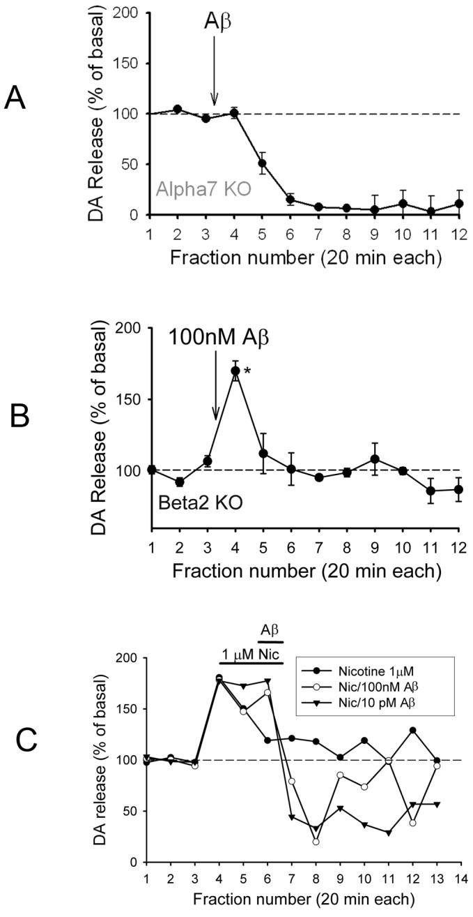
β-amyloid-evoked DA outflow in prefrontal cortex of mice harboring null mutations of nAChR subunits. (A) Perfusion with 100pM or 100nM Aβ1-42 for 5min into prefrontal cortex of α7 nAChR null-mutant mice (Alpha7 KO). (B) Perfusion with 100nM Aβ1-42 into prefrontal cortex of β2 nAChR null-mutant mice (Beta2 KO). Shorter perfusion with Aβ in A and B was performed in order to reveal possible slowly developing inhibition of DA outflow resulting from the fall-off in Aβ following perfusion. (C) Perfusion with 1μM nicotine for 30min, then 100nM or 10pM Aβ1-42 with nicotine for an additional 20min. Experiments were performed as described in the legend to Fig. 2. Data are presented as averages ± s.e.m; n=3-5 replicates each, except in C where only averages are shown for clarity (n=3). Mean basal values for DA: 0.5-1pg/ul, adjusted for recovery (8-23%); *p<0.05 relative to baseline.