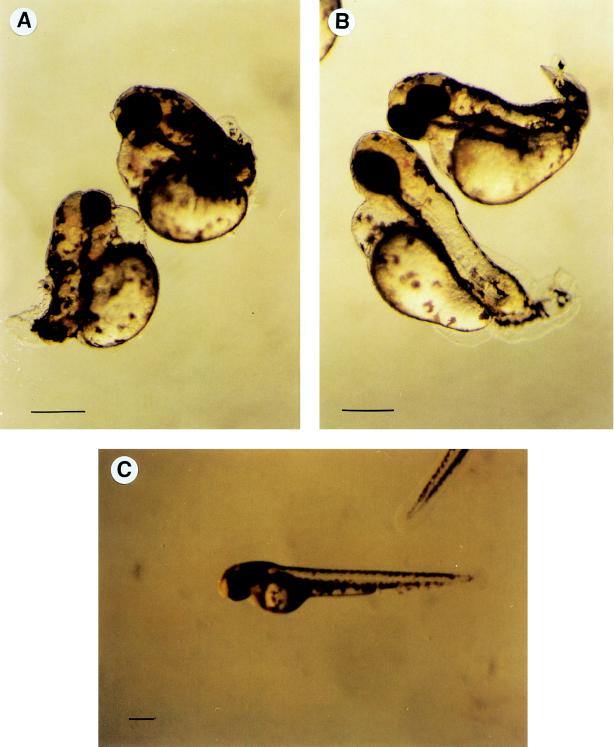
Figure 3.
Results of microinjection experiments in zebrafish. All embryos shown are 48 h old and represent typical examples. (A) Embryos injected with DG42 antiserum. Sixty-one percent of the embryos (n = 288) were consistently and reproducibly affected in the formation of the trunk and tail. (B) Embryos injected with NodZ protein. Sixty-nine percent of the injected embryos (n = 106) show defects similar to those observed after injection of the DG42 antiserum. As a control, embryos were injected with an identical preparation of NodZ protein inactivated prior to injection by boiling for 5 min. Sixteen percent of these controls (n = 98) were affected, although the defects observed are nonspecific and do not resemble those seen when injecting the active protein (data not shown). (C) Control embryos injected with rabbit preimmune serum. Five percent of control embryos (n = 116) were affected by the injection procedure, but the observed defects were not specific. (Bars = 250 μm.)

