Fig. 1.

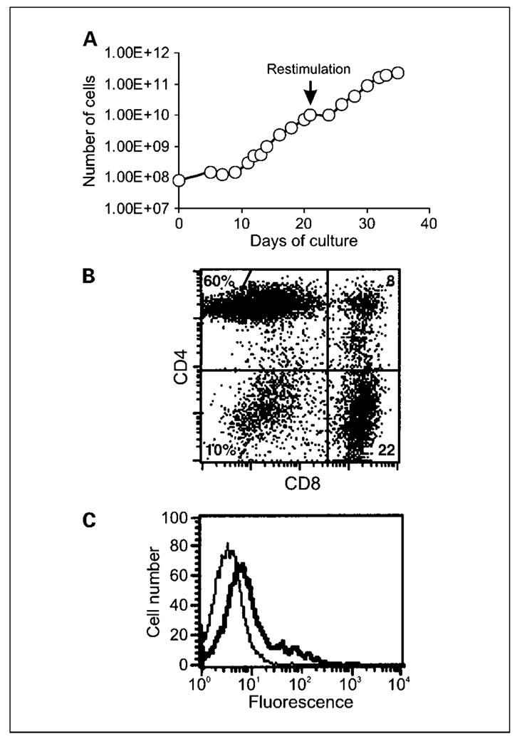
Growth and phenotype of gene-modified T cells. A, following allogeneic stimulation, 8 × 10⁷ Tcells were transduced with retroviral vector encoding the MOv-γ receptor and maintained at 1to 2 × 10⁶/mL in media containing IL-2. Transduced T cells were restimulated with allogeneic PBMCs on day 21, which resulted in further expansion of Tcells. Using this method, large numbers of dual-specificTcells could be generated. T cell expansion depicted is for patient 9. Representative of all six patients in cohort 2.The phenotype of transduced Tcells from cohort 2 of the study was determined with respect to T cell subset markers and chimeric receptor expression using specific antibodies and flow cytometry. B, although the relative proportions of CD4+ and CD8+ T cells varied between patients, the culture was made up predominantly of CD4+ and CD8+ cells as seen in the representative plot. C, expression of the chimeric MOv-γ receptor was evident following staining with anti-idiotype antibody (thick line) compared with isotype control antibody (thin line). Representative of all patients.
