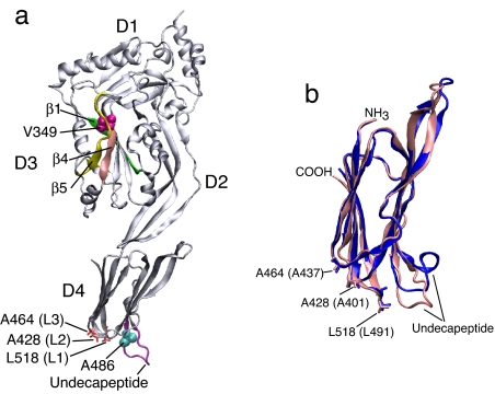
Fig. 1.
The crystal structure of ILY and the domain-4 crystal structures of ILY and PFO. (a) A ribbon representation of the crystal structure of ILY (34) denoting the positions of various structures and residues referred to in this work. (b) An overlay of a ribbon representation of the D4 structures of ILY (pink) and PFO (blue) based on the crystal structures of both proteins (34, 44). Shown are the locations of the undecapeptide and the L1–L3 loop residues of ILY and PFO (the PFO loop residues are in parentheses). The images were generated by using Visual Molecular Dynamics (45).

