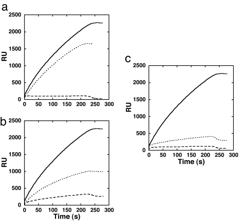
Fig. 4.
The L1–L3 loops mediate PFO binding to cholesterol-rich liposomes. Shown is SPR-binding analysis of aspartate- and glycine-substituted PFO loop mutants for residues Ala-401 (loop L2), Ala-437 (loop L3), and Leu-491 (loop L1). (a) SPR-detected binding of native PFO (solid line), PFOA401D (dashed line), and PFOA401G (dotted line). (b) SPR-detected binding of native PFO (solid line), PFOA437D (dashed line), and PFOA437G (dotted line). (c) SPR-detected binding of native PFO (solid line), PFOL491D (dashed line), and PFOL491G (dotted line). RU, resonance units.

