Fig. 2.
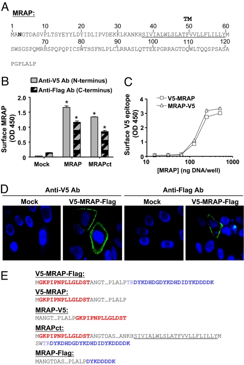
Transfected MRAP is inserted in the membrane in two opposite orientations. (A) Amino acid sequence of mouse MRAP. The underlined sequence is the predicted transmembrane domain, and the Asn shown in bold is the single natural potential N-linked glycosylation site. (B) Detection of both V5- and Flag-epitopes on the surface of nonpermeabilized CHO cells expressing V5-MRAP-Flag or MRAPct with the same tags measured by ELISA. (C) Detection of surface V5-epitope by ELISA in CHO cells expressing V5-MRAP or MRAP-V5. (D) Surface staining of live CHO cells expressing V5-MRAP-Flag with anti-V5 (Left) or anti-Flag (Right) antibodies (shown in green); nuclei are counterstained in blue. (E) Epitope-tagged MRAP constructs. V5 epitopes are shown in red, and Flag epitopes are shown in blue.

