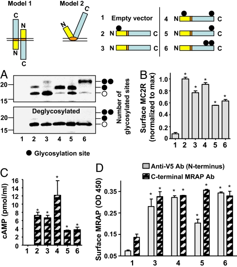
Fig. 4.
MRAP spans the membrane in two orientations. (A) CHO cells transfected with different glycosylation mutants of V5-tagged MRAP were lysed and MRAP immunoprecipitated with anti-V5 antibody and deglycosylated or not with PNGaseF. Blots were probed with anti-V5 antibody. When surface MRAP was selectively isolated (as described for Fig. 5B), approximately equal amounts of glycosylated and nonglycosylated MRAP were seen with MRAP-N3Q/Q96N/P88T. (B–D) Glycosylation mutants of MRAP are functional and display dual topology. CHO cells were transfected with empty vector, HA-MC2 receptor, and wild-type or V5-tagged mutant MRAP constructs. (B) Surface ELISA of HA-MC2 receptor (MC2R). (C) cAMP response to 100 nM ACTH. (D) Surface ELISA of N-terminal MRAP detected with anti-V5 antibody and C-terminal MRAP detected with affinity-purified rabbit anti-C-terminal MRAP antibody. *, P < 0.05 versus mock-transfected.

