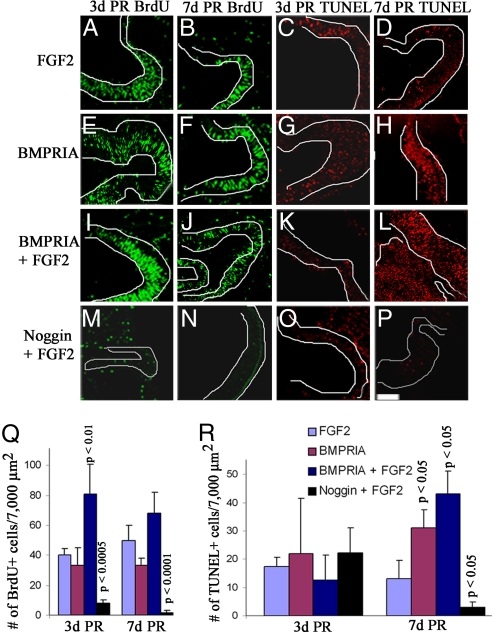
Fig. 3.
Activation of BMPRIA induces proliferation during early stages of retina regeneration and apoptosis during late stages of regeneration. (A–P) Retinectomies were performed on E4 chick eyes and collected at 3d PR (A, C, E, G, I, K, M, and O) or 7d PR (B, D, F, H, J, L, N, and P). Eyes were treated with FGF2 (A–D), RCAS BMPRIA (E–H), RCAS BMPRIA + FGF2 (I–L), or RCAS noggin+ FGF2 (M–P). (A, B, E, F, I, J, M, and N) Immunohistochemistry by using an antibody for BrdU (green) shows BrdU+ cells in the anterior region during retina regeneration at each stage. (C, D, G, H, K, L, O, and P) TUNEL analysis shows cells undergoing apoptosis at each stage. (Scale bar: 200 μm; scale bar in P is for all images.) (Q and R) Graphical representation of the average number of BrdU+ (Q) or TUNEL+ (R) cells at each stage in each treated group. P values represent significance, compared with eyes treated with FGF2 at each stage. The key shown applies to both graphs.

