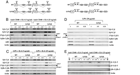
Fig. 3.
Analysis of GL transcription. (A) Relative position of some primers used for RT-PCR on the spliced (dotted line) γ3 and γ1 transcripts (Left) and on unspliced transcripts (Right) from WT and Iγ3/Iγ1 alleles respectively (not to scale). (B) RT-PCR was performed on WT or Iγ1/Iγ3 GL transcripts from anti-CD40+IL4-activated splenocytes RNA (day 3) for Iγ1-Cγ1, Iγ3-Cγ3, Iγ2b-Cγ2b, 3′Iγ1-Cγ3/1 GL, or AID and actin transcripts, respectively. Single-stranded cDNAs or dilutions thereof (1/5 and 1/25) were subjected to PCR, using appropriate primers. Arrows indicate transcripts initiating from the endogenous Iγ1 promoter (upper band, referred to as γ1 GLT) or from the replacement promoter (lower band, γ1/γ3 GLT). (C) RT-PCR was performed as in A on GL transcripts from LPS+IL4-activated splenocytes. (D) RT-PCR was performed as in A on GL transcripts from LPS-activated splenocytes. (E) RT-PCR was performed on WT or Iγ1/Iγ3 RNAs from nuclei of anti-CD40+IL4-activated splenocytes for γ1, γ3, γ2b, or β-actin unspliced transcripts, respectively. Single-stranded cDNAs or dilutions thereof (1/5 and 1/25) were subjected to PCR, using appropriate primers.

