Fig. 5.
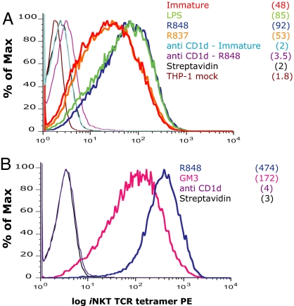
Direct detection of iNKT cell ligand(s) by soluble iNKT TCR staining. (A) THP-1 CD1d cells were activated for 36 h with LPS, R-848, or R-837. The presence of iNKT cell ligands on the surface of THP-1 CD1d cells was revealed by staining with the soluble iNKT TCR in the presence of isotype control or blocking anti-CD1d antibodies. (B) THP-1 CD1d cells were activated for 24 h with R-848 and incubated with an excess of the ganglioside GM3 overnight or blocking anti-CD1d antibodies before staining with the soluble iNKT TCR. Streptavidin staining is included as negative control. THP-1 mock transduced cells and DCs did not show any detectable staining (data not shown). Numbers in brackets represent the mean fluorescence intensity (MFI) values of the iNKT TCR staining. CD1d MFI remained unchanged with maturation (data not shown). One experiment representative of six is shown.

