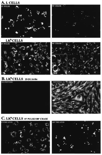
Figure 1.
Internalization of peptides in L and LKb cells. (A) L or LKb cells were exposed (Left) to the KbFL peptide for 15 min and examined by laser scanning confocal microscopy or incubated (Right) for an additional 90 min at 37°C after removal of peptide before examination. (Lower Right, Inset) Enlargement of the region around the cell marked with an arrow to better observe the reticular staining pattern in the cytoplasm. (B) LKb cells were treated with both 40 units/ml γIFN and 1 μg/ml BFA/ml for 2 h and 10 μM cbz-LeuLeuLeu (generously provided by M. Orlowski, Mt. Sinai School of Medicine, New York) for 1 h before addition of 10 μg/ml unmodified SIINFEKL peptide for 1 h. Cells were stained with fluorescein conjugated-25-D1.16. (C) LKb cells were incubated for 15 min with KbFL and then for 180 min at 37°C in the absence (Left) or presence (Right) of aluminum fluoride.

