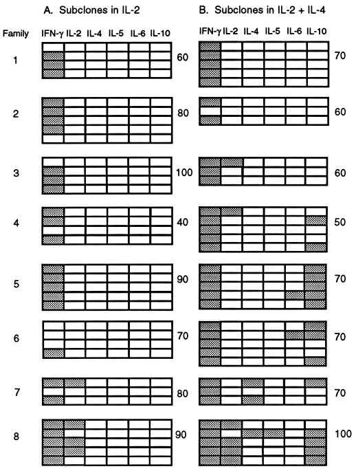
Figure 4.
Cytokine mRNA expression patterns within families of subclones derived after ≥7 cell divisions. Primary clones were grown for 5 days with anti-receptor antibodies and IL-2 and those of 200–300 cells identified microscopically. Twenty individual cells from each clone were transferred into secondary cultures, 10 with IL-2 (A) and 10 with IL-2 + IL-4 (B). After 3 days, RNA was extracted from the five largest subclones of each group (mean 37 ± 31, range 2–130 cells) and assayed for β-actin and cytokine mRNAs. Results are shown for all β-actin+ subclones in eight families. Detection of a cytokine PCR product is indicated by shading of the boxes. Subcloning efficiencies (%) for each group of 10 transferred cells are shown at the right.

