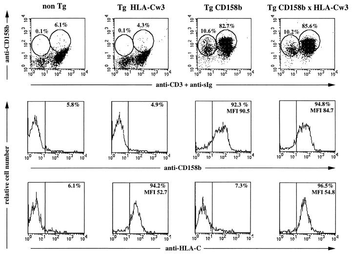
Figure 2.
Cell surface expression of the CD158b transgene. PBL isolated from mice representative of the indicated mouse lines were examined by flow cytometry for the cell surface expression of CD158b, CD3ɛ, surface immunoglobulin (sIg), and HLA-Cw3; nontransgenic, non-Tg; HLA-Cw3 transgenic, Tg HLA-Cw3; CD158b transgenic, Tg CD158b (L61); HLA-Cw3 and CD158b transgenic, Tg CD158b × HLA-Cw3. Cells were stained with FITC-goat anti-mouse IgG; after saturation of free binding sites with mouse Ig, FITC-anti-CD3ɛ and biotinylated GL183 (anti-CD158b) mAbs were added. Biotinylated GL183 was revealed using TC streptavidin. For HLA-Cw3 expression cells were incubated with F4/326 mAb (anti-HLA-C) followed by a FITC-goat anti-mouse IgG. Percentage of positive stained cells in each circle is indicated. (Middle and Bottom) Percentage and mean of fluorescence intensity of CD158+ and HLA-Cw3+ cells are indicated.

