
Figure 6.
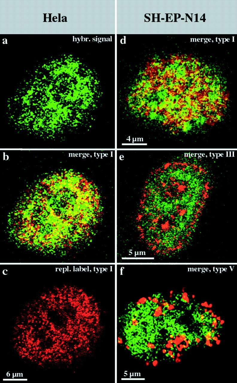
R-band DNA is confined to the interior compartment during interphase. Nuclei (a–c, HeLa S6 nucleus; and d–f, three different nuclei from SH-EP N14 cells) were replication-labeled with Cy3-dUTP (replication patterns depicted in red). DNA from the H3 isochore fraction (FITC-detected, depicted in green) was hybridized to Cy3-labeled nuclei. Nuclei were analyzed by confocal microscopy. For each nucleus, identical midnuclear planes are shown regarding FITC or Cy3 fluorescence detection (colocalizing FITC and Cy3 fluorescence appears yellow on merged [b and d–f] images). Regarding the HeLa nucleus, the merge (b) of the hybridization signal (a) and the type I replication pattern (c) reveals the localization of R-band sequences within the interior compartment. A similar localization of R-band sequences is obvious regarding SH-EP N14 cells (d, underlying type I pattern in red). R-band sequences are excluded from the peripheral (e, red, type III pattern) and late replicating (f, red, type V pattern) compartments. It should be noted that all confocal images presented were not further processed and reflect the resolution limits of light microscopy. Therefore, some colocalization of fluorescent signals at the boundaries of differently labeled distinct compartments is expected.