
Figure 10.
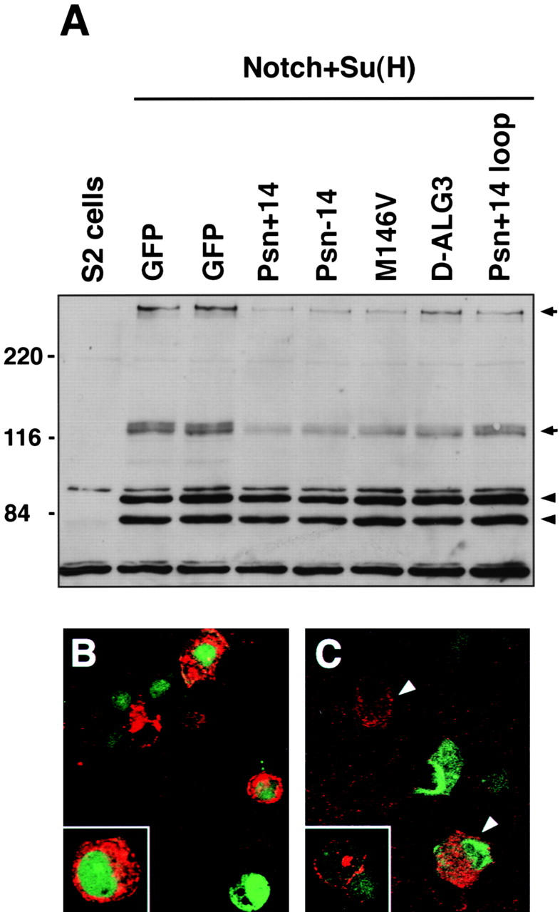
Expression of Psn leads to reduced Notch protein levels in S2 cells. A, S2 cells were transfected with pMTNMg, which expresses full-length Notch; pHS-Su(H)4, which expresses Myc-tagged Su(H); and a pair of overexpression constructs consisting of pHS-GV, which expresses a GAL4-VP16 fusion protein, along with either pUAS-Psn or pUAS-GFP constructs, as labeled above each lane. Cell lysates were analyzed by immunoblotting with a mixture of three antibodies: mAb C17.9C6, which detects both the ∼300-kD full-length and ∼120-kD COOH-terminal Notch fragments (arrows); mAb 9E10, which detects the Myc-tagged Su(H) doublet bands (arrowheads); and mAb E7, which detects β-tubulin (at bottom of gel). Both full-length and COOH-terminal Notch fragment levels are significantly reduced when the Psn constructs are expressed before Notch expression, whereas levels of Su(H) and tubulin are unaffected. B, S2 cells transfected with pMTNMg were fixed and stained with mAb C458.2H, an antibody recognizing the extracellular domain of Notch. In pMTNMg-transfected cells (marked by green GFP cotransfection), Notch accumulates at high levels (red) and shows both vesicular cytoplasmic and cell-surface staining (inset, higher magnification view). When Psn is coexpressed in these cells (C), Notch levels are dramatically reduced (arrowheads). No obvious vesicular staining is detected in these cells, although Notch is still normally localized in the plasma membrane, as shown by cell-surface antibody staining of live, nonpermeabilized S2 cells (inset).