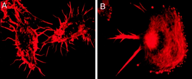
Figure 5.
F-actin distribution in the two macrophage cell lines by epifluorescence microscopy. (A) Nonmotile P388D1 cells stained with rhodamine phalloidin show F-actin staining in filopodia, cell surface microvilli and the cell body. (B) Staining of motile IC-21 cells with rhodamine phalloidin detected F-actin in clusters of small adhesions at the leading edge of the cell, in the cell body, and in retraction fibers at the trailing end of the cell.

