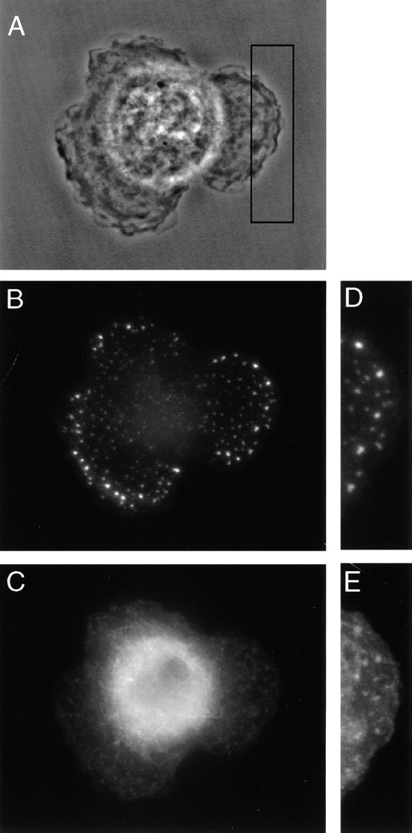
Figure 7.

Localization of the fimbrin–vimentin complex in spreading cells by phase and epifluorescence microscopy. Half hour after plating, IC-21 cells are seen spreading out in all directions (A). Fimbrin (B and D) and vimentin (C and E) are detected in common foci in regions of the cell that are actively spreading. The vimentin network is collapsed around the nucleus.
