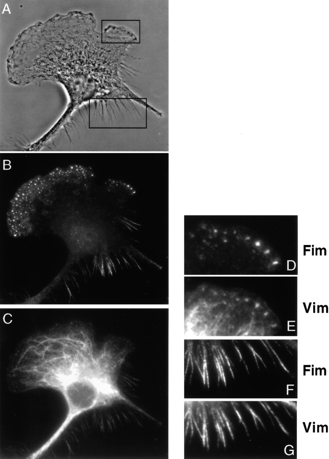
Figure 8.
Localization of the fimbrin–vimentin complex in polarized IC-21 cells. 3 h after attachment, the cells show, by phase microscopy, an asymmetrical morphology (A). Using epifluorescence microscopy the fimbrin–vimentin complex is shown to be restricted to foci present at the cells leading edge where the lamella is actively expanding (D and E). The vimentin network is extended out in the direction of the leading edge (C). The fimbrin–vimentin complex is also detected in retraction fibers at the trailing end of the cell (F and G).

