Figure 6.

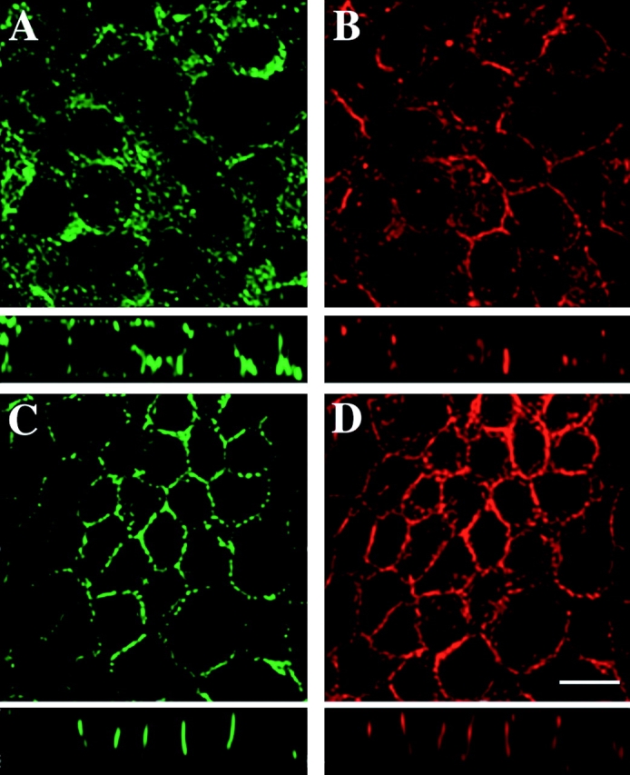
Stabilization of actin by phalloidin prevents redistribution of CD44 upon digitonin-induced depletion of plasma membrane cholesterol. Extended focus images and vertical (x-z) sections are shown. In digitonin-permeabilized EpH4 cells, CD44 stained by the respective antibody was redistributed to the apical surface (A; similar to the experiment shown in Fig. 5). The actin cytoskeleton is visualized by phalloidin-rhodamine, failing to show an extensive subcortical actin ring in many cells (B). When phalloidin was added to the permeabilization buffer (see Fig. 5), CD44 remained precisely localized to the basolateral plasma membrane domain (C). Phalloidin-rhodamine showed enhanced staining of filamentous actin and stabilization of the subcortical actin ring upon treatment with phalloidin (D). Bar, 10 μm.
