
Figure 2.
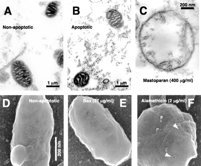
Apoptotic mitochondria exhibit normal size and morphology on electron microscopy. (A–C) Crude extracts were incubated at 0°C as a non-apoptotic control (A) or incubated at 22°C to induce spontaneous cytochrome c release (B). Immediately after cytochrome c release in the sample taken at 2.75 h, aliquots were fixed and examined by transmission electron microscopy. (C) A swollen mitochondrion taken from a further sample treated with mastoparan (400 μg/ml). (D–F) FEISEM of reconstituted extracts incubated at 0°C for 3 h, 40 min (D, non-apoptotic) or 22°C in the presence of 35 μg/ml BaxΔTM for 1 h, 40 min (E). (F) Alamethicin addition (2 μg/ml, 40 min) caused both cytochrome c release and the formation of 10–13-nm pores (arrows). Bar, (D–F) 200 nm.