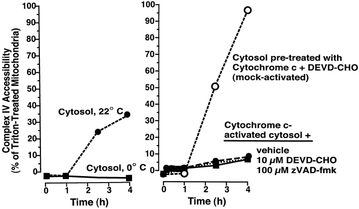
Figure 9.
PEF is inactivated by caspases. Mitochondria were incubated with either normal cytosol at 0 or 22°C (left), or with cytosol that had been activated by preincubation (3 h) with 0.5 μM horse heart cytochrome c (HHCc) (right). Samples in activated cytosol contained either vehicle (0.5% DMSO), Ac-DEVD-CHO (10 μM) or zVAD-fmk (100 μM). One sample (mock-activated), had Ac-DEVD-CHO (10 μM) added before incubation with HHCc. At the indicated times, aliquots (2 μl) were assessed for complex IV accessibility. Mitochondria in the three activated cytosol samples had lost their cytochrome c by 1 h due to tBid-like activity (not shown). Data are representative of three experiments.

