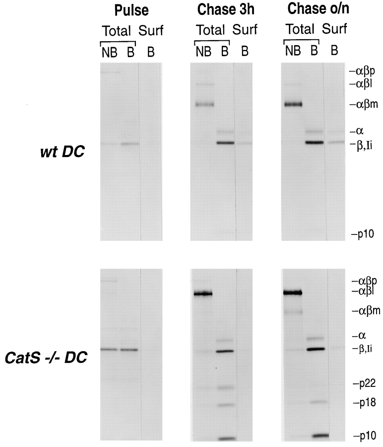
Figure 8.
Surface labeling of MHC class II molecules in DC from wt and CatS−/− cells. Flt3-induced DC from wt (upper panels) and CatS−/− (lower panels) mice were pulsed with [35S]cysteine/methionine for 60 min, and chased 3 h and overnight (o/n), respectively. At each chase point the cell surface was biotinylated as described. The samples were immunoprecipitated with N22 to retrieve total cellular MHC class II. 10% of this immunoprecipitate was divided into two and loaded on a SDS-PAGE without or with prior boiling (total, NB, and B). The remaining 90% were reimmunoprecipitated with streptavidin-agarose beads, extensively washed, and analyzed on the same 12.5% SDS-PAGE after boiling (surf, surface; B, boiled).

