Figure 3.
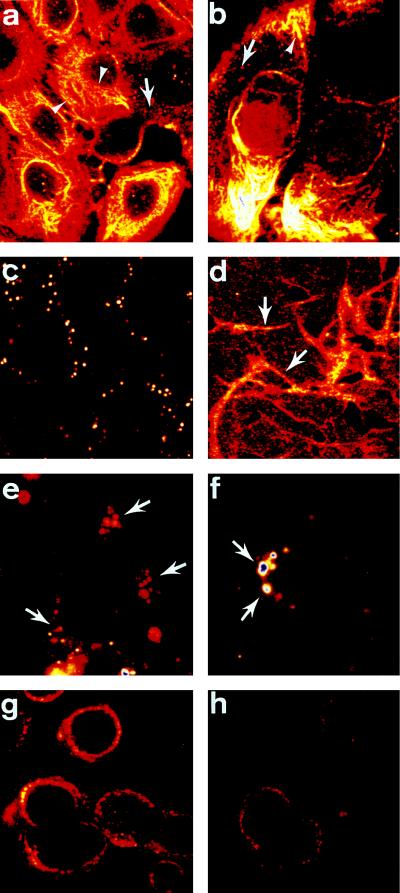
COU-1 localization, internalization, and influence of fibronectin on surface cytokeratin expression. Colon cancer cells [H3619 (a), colo137 (b)] were fixed, permeabilized, and stained with COU-1 (a) or Fab COU-1 (b) and FITC-labeled goat anti-human κ-chain antibody. Note intense fibrillar staining of the intermediate filament (arrowhead). In addition, vesicles (arrows) throughout the cytoplasm were routinely observed. Live H3619 cells incubated with COU-1 at 4°C gave dispersed punctate staining of the upper cell surface (c). When the H3619 cells were grown on fibronectin-coated slides, the level of punctate staining at the cell surface increased and COU-1 was enriched at intercellular junctions (arrows, d). When the colon cancer cells [H3619 (e, f), colo137 (g, h)] were incubated with COU-1 at 37°C, the dispersed punctate surface staining disappeared and COU-1 (e, f, h) or Fab COU-1 (g) localized in large cytoplasmic vesicles adjacent to the nucleus. (×1000.)

