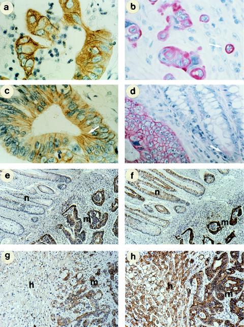
Figure 4.
Comparison of the tissue distribution of COU-1 and murine anti-cytokeratin 8 antibody in ethanol-fixed tissues. Tumor cells within tissue sections bound COU-1, while surrounding normal cells were not stained (a–d). Fibrillar staining characteristic of intermediate filaments (a, arrow), membrane staining of single proliferating cells (b, arrow; note metaphase, arrowhead), and enrichment of COU-1 at intercellular junctions (arrows in c and d). In adjacent normal colon epithelia, weak staining was found only in a few cells of some biopsies (arrow, d). (e and f) Comparison of staining with COU-1 (e) and murine anti-cytokeratin 8 (M20) (f) on serial sections of malignant (m) and adjacent normal colon epithelium (n). (g and h) Adjacent sections of a colon cancer metastasis in the liver (m) and surrounding normal hepatocytes (h) incubated with COU-1 (g) and with M20 (h). (a and b, ×600; c and d, ×500; and e–h, ×200.)

