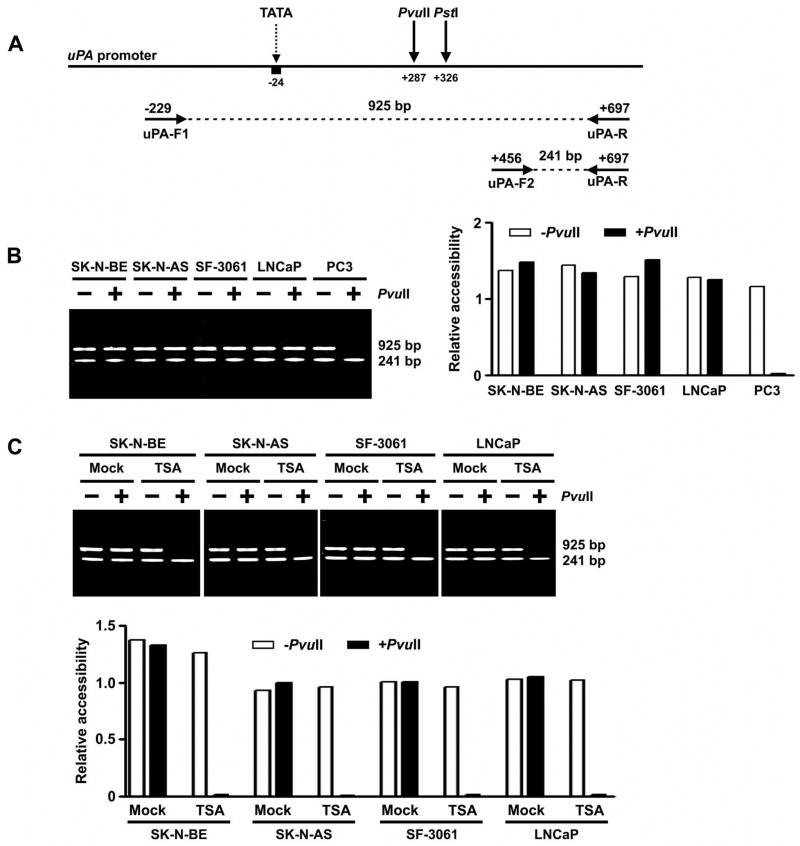
Figure 3. Chromatin conformation of the uPA promoter as determined by restriction enzyme accessibility assay.
A. Strategy used to perform restriction enzyme accessibility assay of the uPA promoter. Genomic DNA of each cell line was digested with restriction enzyme PvuII before PCR amplification with a set of three primers (uPA-F1, uPA-F2, and uPA-R). The ratio of 925:241 bp products reflects promoter accessibility.
B. Left, RE accessibility of the uPA promoter region in human cancer cell lines SK-N-BE, SK-N-AS, SF-3061, LNCaP and PC3. Right, densitometry of undigested (− PvuII) and digested (+PvuII) PCR products. The decrease of the ratio 925:241 bp after PvuII digestion indicates that the chromatin is accessible to PvuII ( p <0.05).
C. Top, chromatin accessibility changes of the uPA promoter region in SK-N-BE, SK-N-AS, SF-3061 and LNCaP cells following treatment with TSA. Bottom, densitometric analysis of undigested (− PvuII) and digested (+PvuII) PCR products in control and TSA-treated cell lines. The ratio was decreased by the treatment of TSA (p <0.05).
(Results are representative of three separate experiments.)

