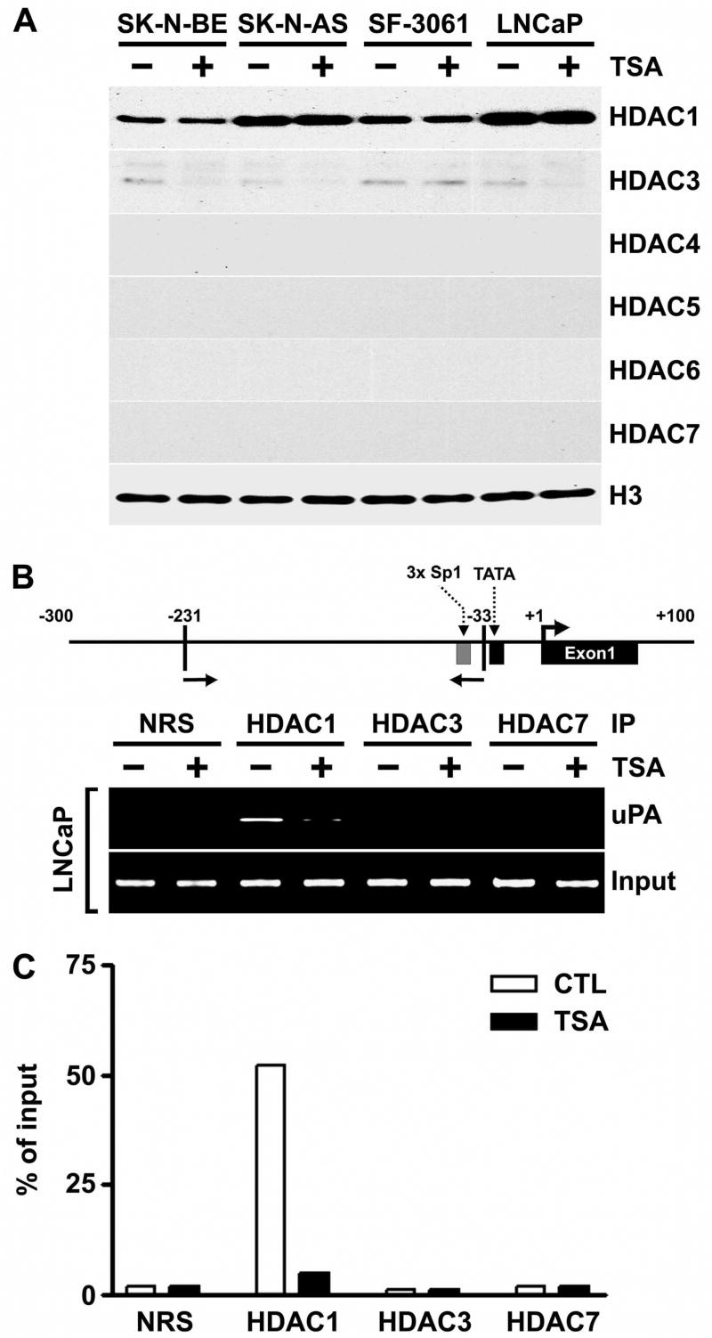
Figure 6. HDAC1 is present in the uPA promoter region in the absence, but not the presence, of TSA.
A. Nuclear extracts were isolated from control and TSA-treated SK-N-BE, SK-N-AS, SF-3061 and LNCaP cells, and immunoblot analysis was performed using anti-HDAC1, anti-HDAC3, anti-HDAC4, anti-HDAC5, anti-HDAC6, anti-HDAC7 and histone H3 antibodies. Histone H3 was utilized as a loading control.
B. LNCaP cells were either treated (+) or not treated (−) with 100 nM of TSA for 8 h and processed for chromatin immunoprecipitation (ChIP) assays. The antibodies used were HDAC1, HDAC3, HDAC7 and control normal rabbit serum (NRS). Top panel: Schematic representation of the uPA promoter, including the locations of the primers used for ChIP. Bottom panel: Immunoprecipitation (IP) inputs and PCR-amplified products resolved on 2% agarose gels and visualized by ethidium bromide staining and UV fluorescence.
C. The ChIP assay was quantitatively determined by ImageJ program, and the results are presented as the percent of the input signal generated with 2% of the chromatin that was immunoprecipitated by the specific antibodies in LNCaP cells.
(Results are representative of three separate experiments.)

