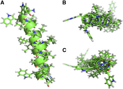
FIGURE 6.
Illustration of proposed WALP23 backbone structure, as viewed from within the membrane interior (A); and viewing the N- and C-termini along the membrane normal (B and C, respectively). Note that residue Leu12 and all side-chain orientations were not determined. This illustration was prepared using Pymol software.

