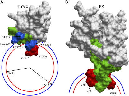
FIGURE 1.
Interaction restraints used for docking the FYVE-DPC and PX-DPC complexes. A and B show the solvent accessible areas of the FYVE and PX structures. Deeply inserting residues (below phosphate groups) are colored in red, interfacial interacting residues (below the micelle surface) are colored in blue, and the additional residues left flexible during docking are colored in green. Visible residues are indicated by arrows. The average positions of the phosphate and choline groups in the DPC micelle (20 and 22 Å from the micelle center) are depicted as red and blue circle sections, respectively.

