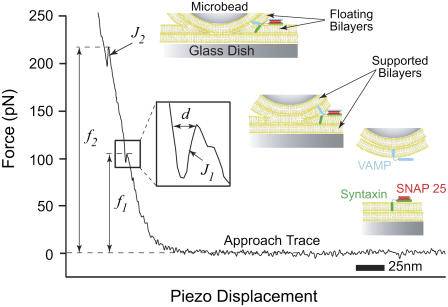
FIGURE 1.
Typical force versus piezo displacement curve displaying the approach trace of an AFM force scan measurement between apposed floating lipid bilayers containing SNAREs. As the bilayers are compressed together, they fuse in two jump steps at J1 and J2. The jump is due to the sudden displacement of the cantilever tip toward the substrate as a result of the merger of the bilayers under the applied compression force f1 or f2. The inset is a magnification of the approach trace during the jump event. Distance d is the measure of the jump distance and is consistently on the order of a single egg PC bilayer thickness. This suggests the merging of the two proximal monolayers (hemifusion) of the apposed bilayers during J1 followed by that of the two distal ones (full fusion) during J2 as depicted in the accompanying cartoons.

