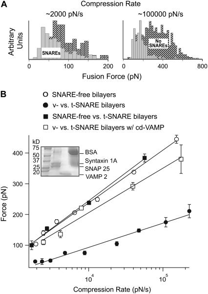
FIGURE 2.
(A) Distribution histograms of fusion force values measured at the first jump during the approach step of an AFM force measurement. The forces measurements were carried out under different compression rates (∼2000 and ∼100,000 pN/s) in the absence (shaded) and presence (hatched) of SNAREs in the bilayers. Notice the shift in the force when SNAREs existed in the bilayers. (B) Dynamic force spectra of the fusion process for egg PC bilayers with and without SNAREs. Notice the significant decrease in the fusion force when cognate v-SNARE (VAMP 2) and t-SNAREs (binary syntaxin 1A and SNAP 25) were present in the bilayers as compared to SNARE-free bilayers. The inset shows sodium dodecylsulfate-polyacrylamide gel electrophoresis confirming the successful reconstitution of VAMP or syntaxin and SNAP 25 in v- and t-SNARE vesicles, respectively. No change in the force profile was observed when v-SNARE bilayers were substituted with SNARE-free bilayers. Similarly, treatment of the t-SNARE bilayers with the soluble cytoplasmic domain of VAMP (cd-VAMP, 20 μM) prevented reduction in the fusion force that was observed for intact v- and t-SNARE bilayers. Lines are fits of the model to the data points. Error bars are the standard error of the mean.

