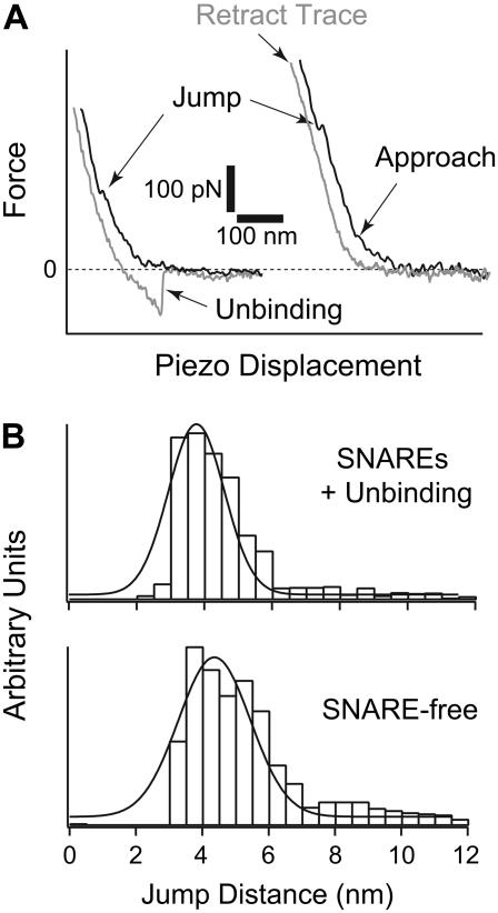
FIGURE 5.
(A) Representative approach and retract traces from AFM force measurement. In the presence of SNAREs in the bilayers (left panel), hemifusion (jump) was observed during approach at a lower compression force, and unbinding events were detected in the retract step. These unbinding events indicated SNARE-mediated fusion. On the other hand, higher compression forces were measured for hemifusion and no unbinding events were detected during the retract step in the absence of SNAREs (right panel). (B) Distribution histograms for jump distance values (d; inset in Fig. 1) measured during jump events in the presence (top) or absence (bottom) of SNAREs in the bilayers. The top panel shows jump distance values that were measured in the presence of simultaneous unbinding events in the retract step. A shift in the jump distance value was observed when SNAREs existed in the bilayers. It was reduced from 4.4 ± 0.1 nm to 3.9 ± 0.3 nm, which is interpreted as an increase in the compressibility and deformation of the membranes when SNAREs are present in the bilayers. The increase in the compressibility and deformation of the membrane translates into a greater ease in membrane fusion.

