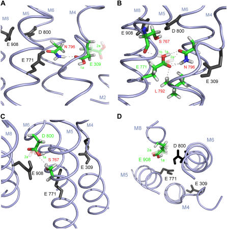
FIGURE 5.
Acidic Ca2+ ligands with hydrogen-bonding partners in the E2( ) state at pH 6. Shown are representative side-chain structures discussed in the text. The respective ligand and interacting residues are colored, other ligands are in black (and without hydrogens except for the hydrogens of the carboxyl groups). For a clearer representation, backbone atoms are not shown, except for Ser767 in Fig. 5 B, since its backbone carbonyl oxygen can be involved in hydrogen bonding to Glu771. Predominantly occupied side-chain structures are solid; less occupied ones are transparent. Black dashes indicate possible hydrogen bonds. M indicates transmembrane helices. In A–C, the cytoplasmic side is on the top of the figure; the lumenal side on the bottom; D shows a top view from the cytoplasmic side. (A) Glu309. Two representative structures are shown: structure 1a (solid) and structure 2a (transparent). The carbonyl oxygen of structure 1a can form a hydrogen bond to Asn796; (B) Glu771. Three representative structures are shown: structure 1a (solid), structure 1b (transparent), and structure 2a (transparent); structures 1a and 1b differ only in hydrogen position. The carbonyl oxygen of all side-chain conformations can form a hydrogen bond to Asn796, only some (structures 1a and 1b) can also form a hydrogen bond to Leu792. The carboxyl hydrogen of some of the side-chain conformations can also form hydrogen bonds to the oxygen of Asn796 (structures 1a and 2a) or Ser767 (structure 1b); (C) Asp800. Two representative side-chain conformations are shown: structure 1a (solid) and structure 2a (transparent). Ser767 can be a hydrogen donor for some Asp800 side-chain conformations (structure 2a). (D) Glu908: Two representative structures are shown: structure 1a (solid) and structure 2a (transparent). More detailed explanations are in the text.

