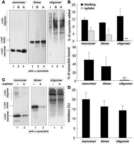
Figure 2. Degradation in lysosomes by CMA of multimeric forms of α-syn.
(A) Association of monomers and irreversibly cross-linked dimers and oligomers of α-syn with isolated lysosomes untreated (binding [B]) or previously treated with proteinase inhibitors (association [A]). Lane 1 shows one-tenth of the amount of protein added to the incubation (input [I]). (B) Upper panel: percentage of each protein bound and translocated (uptake = association – binding) inside lysosomes, calculated from the densitometric quantification of 6–8 immunoblots as used for the immunoblots shown in A. Lower panel: percentage of α-syn bound to the lysosomal membrane that is translocated into lysosomes. (C) Effect of a 2-molar excess of GAPDH on the association of monomeric, dimeric, or multimeric α-syn with lysosomes. (D) Percentage of inhibition of the lysosomal association of each form of α-syn calculated from the densitometric quantification of 4–6 immunoblots such as those shown in C. **P < 0.01.

