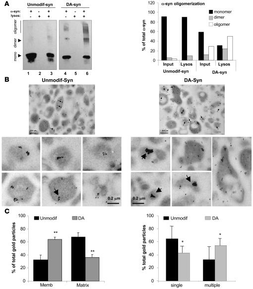
Figure 7. Organization of dopamine-reacted α-syn at the lysosomal membrane.
(A) Association of unmodified (Unmodif-syn) and dopamine-reacted α-syn (DA-syn) to isolated lysosomes was analyzed after incubation by SDS-PAGE and immunoblot for α-syn. Monomers (mono), dimers, and oligomers are indicated by arrows. Lanes 1 and 4: one-tenth of the protein added to the incubation (inputs); lanes 2 and 5: lysosomes incubated alone; lanes 3 and 6: lysosomes incubated with α-syn. Right panel: percentage of α-syn in each of the different multimeric states in the input and associated with lysosomes was calculated by densitometric analysis of the immunoblots. (B) Electron microscopy and immunogold with an antibody against α-syn of lysosomes incubated as in A. The contribution of nonlysosomal structures to this fraction was less than 0.01%, and the percentage of total lysosomes active for CMA in this fraction was 95%. Arrows indicate clusters (>5) of gold particles. Panels on the bottom show individual lysosomes at higher magnification to better show the size of the gold particle clusters. (C) Percentage of gold particles associated with the lysosomal membrane and matrix (left panel) and present as single particles or organized in clusters (>5 gold particles) (right panel) in lysosomes incubated with unmodified and dopamine-reacted α-syn. Values are mean + SEM of the quantification of 4 fields (approximately 150 lysosomes). *P < 0.05; **P < 0.01.

