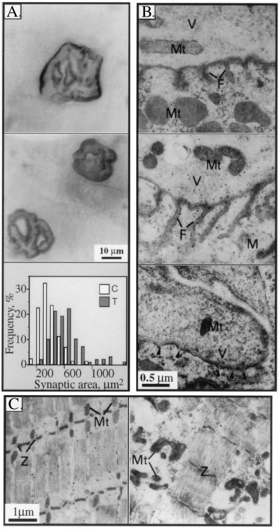
Figure 6.
Enlargement and shape modifications in diaphragm neuromuscular junctions of transgenic animals. (A) NMJ surface changes. Wholemount cytochemical staining of AChE activity was performed on fixed diaphragms from 4-month-old transgenic (Top) and control (Middle) animals. Transgenic NMJs displayed larger circumference and fading boundaries as compared with the complex, sharp contours in controls. Representative endplates from 12 control and transgenic mice. Similar results were obtained using methylene blue staining for total proteins (not shown). (Bottom) Stained areas in μm2 for motor endplates illustrated above (for 90 control, 100 transgenic terminals) are presented as a frequency distribution plot. (B) Postsynaptic fold changes. Electron microscopy of 80-nm crosssections of terminal diaphragm zones reveals variable deformities in NMJ from transgenic mice. (Top) Normal NMJ. (Middle) Transgenic NMJ with exaggerated postsynaptic folds. (Bottom) Transgenic NMJ with short, undeveloped folds marked by arrowheads. V, vesicles; M, muscle; F, postsynaptic folds. (C) Muscle abnormalities. (Left) Longitudinal section from normal diaphragm muscle. Note the organization of mitochondria (Mt) at well-aligned Z bands (Z). (Right) Transgenic diaphragm muscle with fiber atrophy, loss of muscle fiber organization, and swelling of mitochondria at disrupted Z bands.

