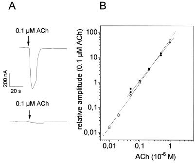
Figure 3.
Effect of DMS cross-linking on ACh-induced current at nAChRs from rat muscle expressed in Xenopus oocytes. (A) Whole-cell current induced by ACh application before (upper trace) and after (lower trace) 30 min of DMS cross-linking (<10% remaining current). The oocytes were washed for at least 10 min after DMS cross-linking, to remove excess of free cross-linker. (B) Dose–response curve before (○) and of the residual current after (•) 30 min of DMS cross-linking (no detectable current after 1 h). The relative amplitude is plotted against the concentration of ACh in a double-logarithmic plot. The amplitudes are normalized to the current induced by 0.1 μM ACh. (After DMS cross-linking it was not possible to record the current below 0.06 μM ACh, because the total current amplitude was below ≈8 nA).

