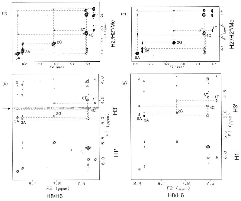
Figure 1.
TRNOE spectra of d(TGACAT) bound to S. cerevisiae Rad51 protein or E. coli RecA protein in the presence of ATPγS. (a and b) d(TGACAT) bound to Rad51 protein. The 0.71 mM d(TGACAT), 67 μM S. cerevisiae Rad51 protein, 0.71 mM ATPγS, 20 mM d11-Tris⋅Cl (pH* 7.1), and 6.7 mM MgCl2 in D2O at 303 K. (c and d) d(TGACAT) bound to RecA protein. The 1.1 mM d(TGACAT), 54 μM E. coli RecA protein, 1.1 mM ATPγS, 20 mM d11-Tris⋅Cl (pH* 7.1), and 6.7 mM MgCl2 and 150 mM NaCl in D2O at 310 K. Mixing time of both spectra is 200 msec. The dotted lines indicate sequential connectivities of d(TGACAT). Signals around 4.7 ppm (indicated by an arrow in b) are derived from residual water.

