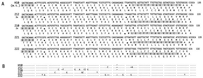
Figure 3.
Sequences of frameshifted H10 mutants (round two). A shows DNA sequences and three reading frames. The FLAG and deduced peptide binder sequences are shaded. Other symbols are as in Fig. 2. B shows a DNA sequence alignment. The alignment compares the H10 sequence with the 221, 222, 212, and 210 sequences. Dots indicate identities, and dashes indicate deletions. The UAG codons are translated as Gln in the figure because the termination of the translation is suppressed in the E. coli strain used.

