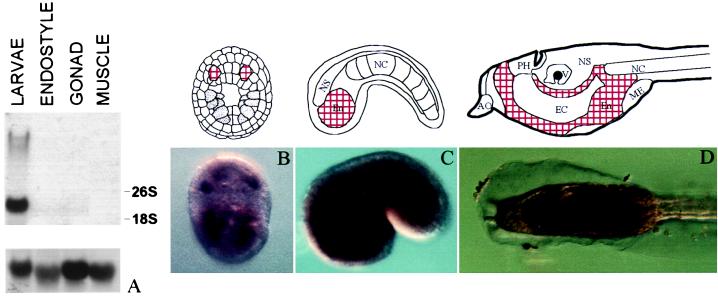
Figure 4.
Expression analysis of CiNR1 in adult and embryonic tissues. (A) Northern blot analysis of CiNR1 in different adult and larval tissues. Ribosomal subunits were indicated. After the removal of the first probe, the filter was hybridized with an oligo against a rat ribosomal subunit (Lower) to test the amount of RNA blotted. (B–D) Whole-mount in situ hybridization of CiNR1 in C. intestinalis. (B) Embryo at neural plate stage; vegetal view. (C) Early tail-bud; lateral view. (D) Swimming larva; lateral view. Each stage is joined to a schematic representation. AO, adhesive organ; PH, primordial pharynx; V, vesicle of prosencephalon; NS, nervous system; NC, notochord; En, endoderm; EC, endodermal cavity; ME, mesodermal pocket. Positive regions are found in the endodermal tissues and correspond to the red areas in the schemes.

