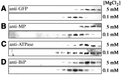
Figure 3.
Western blot analyses of fractions from sucrose density gradients. Membrane preparations with bound (5 mM MgCl2) or displaced (0.1 mM MgCl2) ribosomes were separated on an 18–55% sucrose gradient. Fractions were analyzed with indicated antibodies. Sedimentation was from left to right. (A) Anti-GFP. (B) Anti-MP. (C) Anti-ATPase. (D) Anti-BiP.

