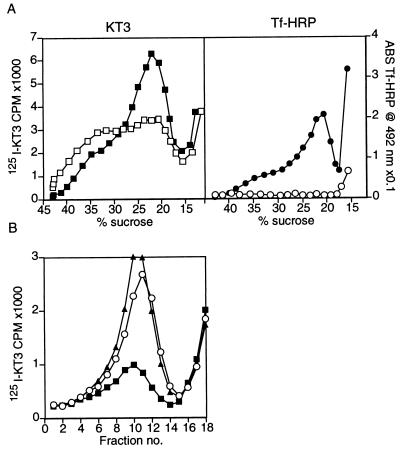
Figure 3.
Tf-HRP is present in the SV precursor endosomes. (A) Endosomes were generated by labeling PC12 cells expressing VAMP-TAg/N49A with both [125I]KT3 and Tf-HRP for 40 min at 15°C, followed by homogenization. 50K g⋅m supernatants were prepared and incubated with DAB and H2O2 for 30 min at 0°C. This caused an HRP catalyzed oxidation of DAB in the Tf-HRP containing organelles and a subsequent crosslinking of their contents. Control reactions were incubated under the same conditions with H2O2 only. Endosomes were then fractionated on sucrose gradients and analyzed for [125I]KT3 and Tf-HRP label. The DAB reaction caused a 53 ± 2% (n = 3) reduction in the KT3 label (Left) appearing at the slow endosomal peak (□), as compared with the control reaction (■). Tf-HRP activity (Right) was completely eliminated after the DAB reaction (○), compared with the control (•). (B) The same endosomes as in A were subjected to an in vitro budding reaction to produce SVs, after the DAB reaction. Membranes were then centrifuged for 35 min at 27,000 × g, and supernatants were analyzed on glycerol velocity gradients. The DAB reaction caused a 67% decrease in budding efficiency (■) as compared with the control (▴) that lacks DAB and H2O2 treatment. This demonstrates that [125I]KT3 is sorted to SVs from a Tf-HRP-containing compartment. When membranes were treated with H2O2 alone (○), no significant decrease in budding efficiency was detected. DAB alone also had no effect on the budding efficiency (data not shown).

