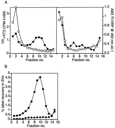
Figure 4.
[125I]KT3 is enriched in slowly sedimenting endosomes and sorted to newly formed SVs. (A) Labeled endosomes in a 50K g⋅m supernatant were incubated under standard reaction conditions at 37°C to generate labeled SVs. The entire reaction mixture was analyzed by sedimentation on a 5 to 25% glycerol velocity gradient, layered over a dense sucrose pad as described in Fig. 1A. A peak of [125I]KT3 label was recovered in fraction 10 (Left) corresponding to newly formed SVs (■). When this in vitro reaction was carried out at 0°C, no SVs were made and the endosome precursors accumulated on the sucrose pad at the foot of the glycerol gradient (□). Tf-HRP label (right) accumulated with the heavy membranes at the bottom of the gradient, whether the in vitro reaction was carried out at 37°C (•), or 0°C (○). This implies that Tf-HRP was not sorted into SVs. (B) Endosomes in a 50K g⋅m supernatant were isolated on sucrose gradients as in Fig. 2. An in vitro reaction was also carried out under standard reaction conditions on another fraction of this 50K g⋅m supernatant. When the reactions were completed, the reaction mixture was subjected to a 35 min centrifugation at 27,000 × g to pellet the donor membranes. After fractionation of the supernatant on glycerol velocity gradients, SVs were identified by a peak of [125I]KT3 in fractions 8–11 (■), characteristic of SVs. No Tf-HRP label was recovered at the SV peak position (•). Plots represent the recovery of label in SVs as a percent of the total label in the endosome donor.

