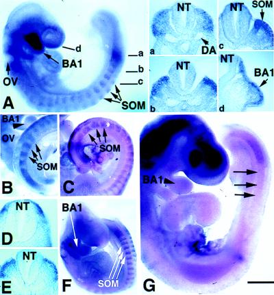
Figure 4.
Distribution of LMO4 mRNA in cranial crest and paraxial mesoderm revealed by whole-mount in situ hybridization. (A) Sixteen somite-stage embryo exhibits LMO4 expression (blue or brown stain) in cranial neural crest migrating into the branchial arches and in rostral somite. Sections at indicated axial levels show expression in dorsal paraxial mesoderm (a), prospective dermomyotome (b), dermomyotome (c), and migratory cranial neural crest in the mandibular component of the first branchial arch (BA1) (d). Otic vesicle (OV), neural tube (NT), and somites (SOM) are indicated by arrows. DA, dorsal aorta. (B) At E10, LMO4 expression in paraxial mesoderm and in rostral somite halves persists at a caudal axial level (E) and is restricted medially at anterior levels to cells adjacent to the neural tube (D). (B) As indicated by arrowhead, LMO4 expression was restricted to ventral mandibular arch by this stage. (C) Pax3 expression is not restricted, but is distributed throughout the dermomyotome. (F) LMO4 activation in Splotch mice is similar to normal embryos in migratory cranial neural crest cells and unsegmented paraxial mesoderm, yet somitic expression is not restricted medially. (G) In Mesp2 mutant embryos, activation of LMO4 in unsegmented mesoderm appears normal. However, expression is lost in rostral somite (arrow). Expression in mandibular arch is detected at a diminished level. Staining in the head region is an artifact caused by probe trapping. [Bar = 200 μm (A, B, C, and G) and 400 μm (D and F).]

