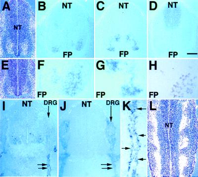
Figure 6.
Activation of LMO4 in motor neurons and Schwann cell progenitors. (A and E) In situ hybridization of the 35S-labeled LMO4 probe to transverse sections counterstained with hematoxylin shows that the domain of LMO4 neural tube (NT) expression (white grains) is broad at a rostral axial level (A) relative to caudal axial level (E). In situ hybridization of digoxygenin-labeled probes to adjacent transverse sections through the E11.5 lumbar spinal cord shows overlap of the LMO4 expression pattern (Blue) (B and F) with motor neuron expression of LHX3 (C and G), but not with LHX3-expressing interneurons. HRP immunohistochemistry shows that the expression domain of the pan-motor neuronal marker HB9 (H) on adjacent sections partially overlaps with LMO4 and LHX3 expression domains. FP, floor plate. F and G are ×2.5 magnifications of B and C, respectively. Apical paraventricular cells that express LMO4 (B) do not overlap with the Pax-3 expression domain (D). (I) At E11.5, activation of LMO4 occurs in cells tightly associated with spinal nerves, indicated by double arrows. (J) Sox10 expression in Schwann cells overlaps with LMO4 expression (I). K is a ×3 magnification of the tissue in I. Arrows indicate individual cells decorating the spinal nerve that express LMO4. (L) Longitudinal section through the E11.5 neural tube hybridized with 35S-labeled LMO4 probe and counterstained with hematoxylin shows expression (white grains) at the exit points for spinal nerves. [Bar = 50 μm (A–E, J), 20 μm (F– H, L), 60 μm (I), and 12 μm (K).]

