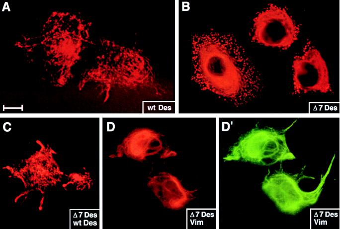
Figure 5.
De novo desmin IF network formation in MCF-7 cells. Cells were transfected with pCVM (not shown), pCMVDes (A and C), pCMVΔ7Des (B–D′), or pJVim (D and D′). After transient expression of the transgenes, cells were fixed and stained with antibodies against desmin (A–C and D′) or vimentin (D). Shown are representative examples of transfected cells. Under these conditions, no staining was observed in pCVM-transfected or in untransfected cells. (Bar represents 15 μm in A and B, 20 μm in C–D′.)

