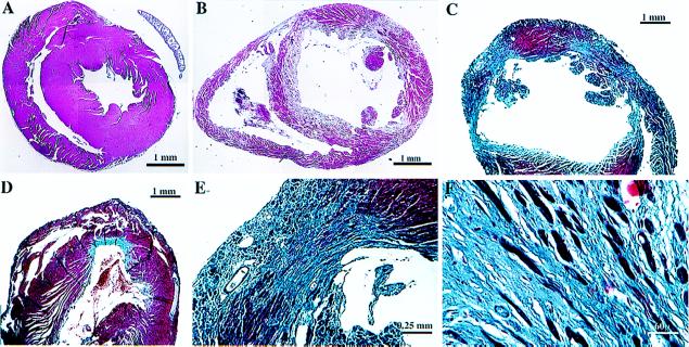
Figure 2.
Histological analysis. Hearts were fixed in formalin and paraffin sections prepared and stained with either hematoxylin and eosin (A and B) or trichrome (C–F) to highlight the interstitial fibrosis. Representative low-power views from a control heart (A) and individual binary transgenic hearts (B–D). Note the chamber dilatation (B and C) and marked variability in the extent of fibrosis (C and D) in binary hearts. Higher power views demonstrate the presence of scant residual myocytes in the most severe lesions (E and F). Scales are indicated. A and B are montages.

