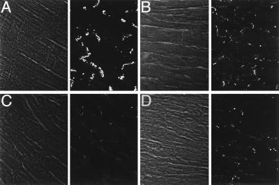
Figure 6.
Cx43 immunostaining. Representative confocal images of Cx43 expression in control (A) and binary transgenic hearts with mild (B), moderate (C), or severe (D) disease. Paired images show phase and immunofluorescence. In the moderately and severely diseased hearts, regions at a distance from focal fibrosis are shown. Images were acquired for equivalent times so that signal intensities could be compared directly. Substantial reduction in the intensity of junctional staining is evident in all transgenic hearts. (Original magnification was 60×.)

