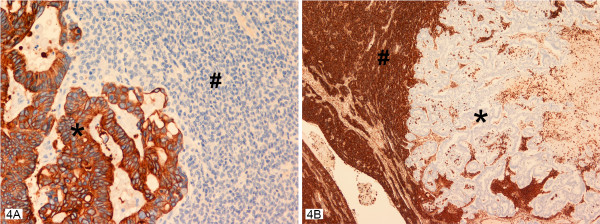
Figure 4.
A, B – Collision tumor with the two neoplastic components seen with their individual staining pattern with cytokeratin 20 antibodies strongly positive in the adenocarcinomatous component (asterisk) in A and negative in the granulosa cell component (pound). While the vimentin antibodies are strongly positive in the granulosa cell component (asterisk) and negative in the adenocarcinomatous component (asterisk) in B. Inhibin staining pattern was identical to that seen with vimentin antibodies.

