
Figure 1.
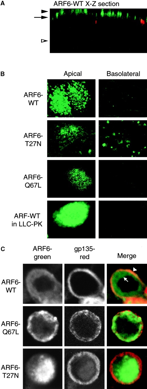
Localization of ARF6 in MDCK cells. A, X-Z section of ARF6–WT: ARF6, green (12CA5 antibody to HA epitope); Z0-1, red. B, X-Y sections taken at AP PM (level of solid arrowhead in A) or BL region (level of open arrowhead in A) of cells expressing indicated protein. ARF6–WT was also expressed in LLCPK1 cells, indicating that the AP localization of ARF6–WT is not unique to MDCK cells. C, X-Y sections taken below apical PM, at level of arrow in A.