Figure 1.

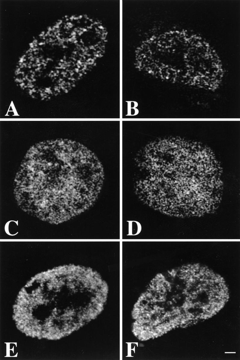
Preservation of the spatial distribution of nascent RNA, DNA, and acetylated histone H4 in human primary fibroblasts during the FISH procedure. Optical sections were obtained before (A, C, and E) and after (B, D, and F) carrying out the FISH protocol. A and B, Optical sections of nuclei in which transcription sites were immunofluorescently labeled. The distribution of nascent RNA did not change significantly during the FISH procedure. C and D, Optical sections of nuclei labeled with the fluorescent DNA stain Sytox green. The pattern of DNA staining did not change significantly after in situ hybridization. E and F, Optical sections of nuclei in which acetylated histone H4 was fluorescently labeled. The distribution did not change significantly during the chromosome painting procedure. The diffuse, low intensity labeling observed before FISH was diminished after the procedure, resulting in a somewhat more pronounced granular labeling. Images shown have been subjected to 3-D image restoration. Bar, 1.75 μm.
